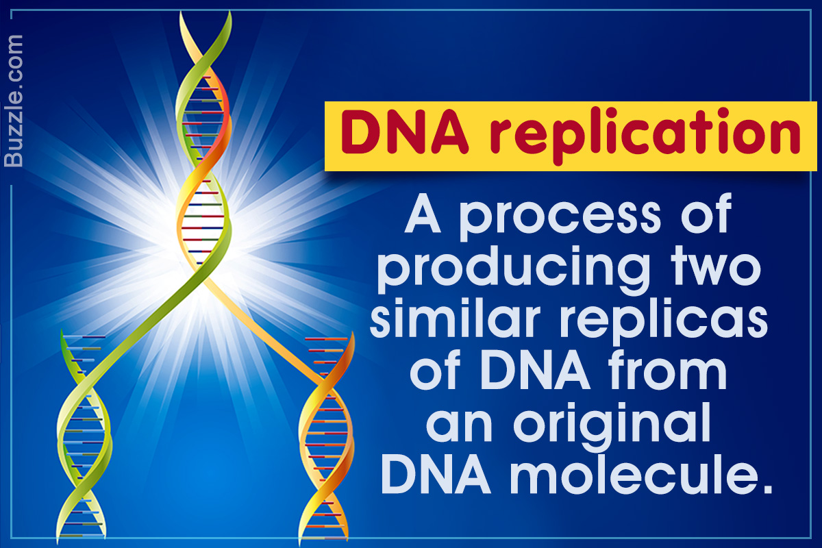
The process of DNA replication comprises a set of carefully orchestrated sequence of events to duplicate the entire genetic content of a cell. The current article provides a short insight into the complex DNA replication steps.
Did You Know?
Less than one mistake occurs while copying a billion nucleotides. This is equivalent to copying 100 dictionaries (with 1000 pages each) word to word, page to page, and symbol to symbol, with only one mistake!
Deoxyribonucleic acid or DNA is the fascinating molecule that stores and passes all the necessary information from one generation to another. It took several interesting experiments by Frederick Griffith, Avery, MacLeod, McCarty, Alfred Hershey, Martha Chase etc., to discover that DNA is the hereditary material. With this foundation, and the research by several scientists like Rosalind Franklin, the structure of this molecule was finally deciphered by James Watson and Francis Crick.
The simple chemical code of the DNA molecule gives rise to the immense complexity of all living organisms. But even more enthralling is its ability to self-replicate and generate another molecule similar to itself. Given below is a brief account of the DNA structure as well as the steps through which a DNA molecule makes copies of itself with an extraordinary accuracy.
Structure of DNA
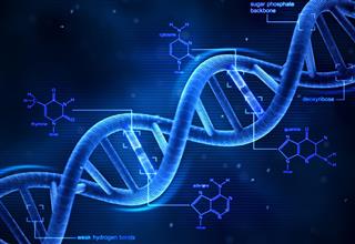
The building blocks of a DNA are molecules called nucleotides, that consists of a deoxyribose sugar (a 5-carbon sugar), a nitrogenous base attached to the sugar, and a phosphate group. There are four types of nucleotide molecules depending on the type of nitrogenous base attached. These four nucleotides (and their respective nitrogenous bases) are:
- Adenosine (Adenine)
- Thymidine (Thymine)
- Guanosine (Guanine)
- Cytidine (Cytosine)
Cytosine and thymine are pyrimidines, a type of six-membered heterocyclic molecules. On the other hand, adenine and guanine are purines, which are two-ringed molecules comprising a pyrimidine ring and an imidazole ring. These nucleotides are linked through their phosphate groups and sugar moieties to form a single strand of the DNA molecule. The phosphate group of one nucleotide and the hydroxyl group of the adjacent nucleotide are connected through a phosphodiester bond. The sugar moieties and the phosphate groups form the backbone of each DNA strand. Each strand has a 5′ phosphate end and a 3′ hydroxyl end.
The DNA double helix comprises two complementary strands that run anti-parallel to each other. One strand runs in the 5′ → 3′ direction, while the other runs in an anti-parallel direction of 3′ → 5′. These strands are attached to each other through hydrogen bonding that occurs between purines and pyrimidines of opposite strands. Adenine pairs with thymine through a double bond (A = T), whereas guanine pairs with cytosine through a triple bond (G ≡ C). The DNA twists at specific lengths due to the bonding angles of the DNA backbone molecules. This forms a helical structure instead of a straight ladder. The A = T and G ≡ C base pairs form the rungs of this helical ladder.
Steps in DNA Replication
The process of DNA replication is a complex one, and involves a set of proteins and enzymes that collectively assemble nucleotides in the predetermined sequence. In response to the molecular cues received during cell division, these molecules initiate DNA replication, and synthesize two new strands using the existing strands as templates. Each of the two resultant, identical DNA molecules is composed of one old and one new strand of DNA. Hence the process of DNA replication is said to be a semi-conservative one.
The series of events that occur during prokaryotic DNA replication have been explained below.
Initiation

DNA replication begins at specific site termed as origin of replication, which has a specific sequence that can be recognized by initiator proteins called DnaA. They bind to the DNA molecule at the origin sites, thus flagging it for the docking of other proteins and enzymes essential for DNA replication. An enzyme called helicase is recruited to the site for unwinding the helices into single strands.
Helicases break the hydrogen bonds between base pairs, in an energy-dependent manner. This point or region of DNA is now known as the replication fork. Once the helices are unwound, proteins called single-strand binding proteins (SSB) bind to the unwound regions, and prevent them for annealing. The replication process thus initiates, and the replication forks proceed in two opposite directions along the DNA molecule.
Primer Synthesis

The synthesis of a new, complementary strand of DNA using the existing strand as a template is brought about by enzymes known as DNA polymerases. In addition to replication they also play an important role in DNA repair and recombination.
However, DNA polymerases cannot start DNA synthesis independently, and require and 3′ hydroxyl group to start the addition of complementary nucleotides. This is provided by an enzyme called DNA primase which is a type of DNA-dependent RNA polymerase. It synthesizes a short stretch of RNA onto the existing DNA strands. This short segment is called a primer, and comprises 9-12 nucleotides. This gives DNA polymerase the required platform to begin copying a DNA strand. Once the primers are formed on both the strands, DNA polymerases can extend these primers into new DNA strands.
The unwinding of DNA may cause supercoiling in the regions following the fork. These DNA supercoils are relaxed by specialized enzyme called topoisomerase which binds to the DNA stretch ahead of the replication fork. It creates a nick in the DNA strand in order to relieve the supercoil.
Leading Strand Synthesis

DNA polymerases can add new nucleotides only to the 3′ end of an existing strand, and hence can synthesize DNA in 5′ → 3′ direction only. But the DNA strands run in opposite directions, and hence the synthesis of DNA on one strand can occur continuously. This is known as the leading strand.
Here, DNA polymerase III (DNA pol III) recognizes the 3′ OH end of the RNA primer, and adds new complementary nucleotides. As the replication fork progresses, new nucleotides are added in a continuous manner, thus generating the new strand.
Lagging Strand Synthesis

On the opposite strand, DNA is synthesized in a discontinuous manner by generating a series small fragments of new DNA in the 5′ → 3′ direction. These fragments are called Okazaki fragments, which are later joined to form a continuous chain of nucleotides. This strand is known as the lagging strand since the process of DNA synthesis on this strand proceeds at a lower rate.
Here, the primase adds primers at several places along the unwound strand. DNA pol III extends the primer by adding new nucleotides, and falls off when it encounters the previously formed fragment. Thus, it needs to release the DNA strand, and slide further up-stream to start the extension of another RNA primer. A sliding clamp holds the DNA in its place as it moves through the replication process.
Primer Removal

Although new DNA strands have been synthesized the RNA primers present on the newly formed strands need to be replaced by DNA. This activity is performed by the enzyme DNA polymerase I (DNA pol I). It specifically removes the RNA primers via its 5′ → 3′ exonuclease activity, and replaces them with new deoxyribonucleotides by the 5′ → 3′ DNA polymerase activity.
Ligation

After primer removal is completed the lagging strand still contains gaps or nicks between the adjacent Okazaki fragments. The enzyme ligase identifies and seals these nicks by creating a phosphodiester bond between the 5′ phosphate and 3′ hydroxyl groups of adjacent fragments.
Termination

This replication machinery halts at specific termination sites which comprise a unique nucleotide sequence. This sequence is identified by specialized proteins called tus which bind onto these sites, thus physically blocking the path of helicase. When helicase encounters the tus protein it falls off along with the nearby single-strand binding proteins.
Fact File
The DNA replication process is almost error free with the help of 3′ → 5′ exonuclease activity of the DNA polymerases. DNA pol III proofreads the nucleotides being newly added to the strand. If a nucleotide has been incorrectly added, DNA pol III recognizes the error immediately, removes the incorrect base, adds the correct nucleotide, and then continues ahead.
Difference Between Prokaryotic and Eukaryotic DNA Replication
Although the basic mechanism remains the same, eukaryotic DNA replication is much more complex, and involves a higher number of proteins and enzymes. The regulatory mechanisms for DNA replication are also more evolved and intricate.
- In prokaryotes, DNA replication is the first step of cell division. On the other hand, eukaryotic DNA replication is intricately controlled by the cell cycle regulators, and the process takes place during the ‘S’ or synthesis phase of the cell cycle.
- Unlike prokaryotic DNA, the eukaryotic DNA is always present in combination with histone proteins that are involved in regulation of gene expression. During replication, these proteins need to be removed just before the unwinding of DNA.
- Owing to higher genomic size and complexity of eukaryotes, several origin and termination sites for replication are present along the DNA. The region between one set of origin and termination sites is called a replication unit or replicon, within which one event of replication takes place. This enables faster and more accurate DNA replication as compared to the prokaryotic system of having a single replicon.
- The Okazaki fragments formed in prokaryotes are longer as compared to those in eukaryotes. In Escherichia coli (E. coli) they are about 1000 to 2000 nucleotides long; whereas in eukaryotes their length ranges between 100 and 200 nucleotides.
- Another interesting difference in prokaryotic and eukaryotic DNA replication is in the termination step of replication. In prokaryotes, the two replication forks, moving in opposite directions along the circular DNA molecule, meet at the termination site, and replication halts. However, eukaryotic DNA being a linear molecule, the lagging strand is shorter than the template strand. To avoid the loss of genetic information through such shortening, chromosomal ends have a set of repetitive sequences called telomeres that comprise noncoding DNA.
Fact File
The human DNA is copied at about 50 base pairs per second. Due to initiation of replication at multiple locations, the process is completed within one hour. If this were not the case, it would take about a month to finish replicating a single chromosome!
The genes of an organism contain all the necessary information to synthesize the right molecule, in the right amounts, and at the right time. Replication is the way to ensure that this coded information is passed down to every cell of the body, and also to the successive generations. After all, as rightly pointed by Richard Dawkins:
“They are the replicators and we are their survival machines. When we have served our purpose we are cast aside. But genes are denizens of geological time: genes are forever.”
