Mitosis and meiosis are two types of cell division processes that play the most crucial role in reproduction, and maintenance of the structural and functional integrity of tissues. Let us understand the various aspects that distinguish these two processes from each other.
Cell theory states that:
- Each living organism is made up of one or more cells.
- Each cell is a collection of organelles dispersed into a membrane bound cytoplasm.
- It is the basic structural, functional and organizational unit of every living organism.
- New cells arise from pre-existing cells through division.
The process of formation of new cells, from the existing ones, can occur through mitosis and meiosis, depending on the cell type and the purpose of division. Given below is a short description of the two processes followed by a detailed account of the differences between them.
Mitosis
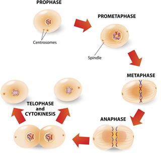
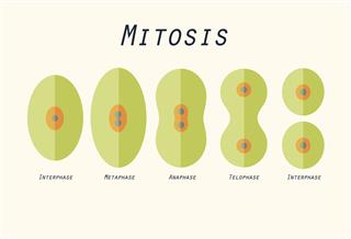
Mitosis is an equational division that involves the duplication of genetic material, and an equal distribution of all the contents into two daughter cells. The cell cycle proceeds via interphase, that comprises the stages of growth and DNA duplication, followed by a mitotic (M) phase. The mitotic phase proceeds through prophase, metaphase, anaphase and telophase. Through these phases, the original nucleus dissolves, the replicated chromosomes align at the center of the cell, and then segregate into two new nuclei. Finally, the cell physically divides into two new cells through cytokinesis.
Meiosis
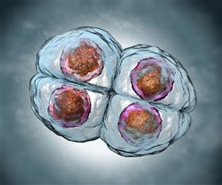
Meiosis is a type of cellular division that results in the formation of four haploid cells from a single diploid cell. During meiosis, the genetic material is replicated only once whereas the nucleus divides twice resulting in ploidy reduction. This is achieved through two successive divisions, meiosis I and meiosis II. The cell cycle events proceed through interphase I, meiosis I, cytokinesis, meiosis II followed by another event of cytokinesis.
Interphase I involves cell growth and chromosome replication. Meiosis I involves the pairing of homologous chromosomes, and their segregation. It is followed by cytokinesis resulting into two haploid daughter cells, but with intact sister chromatids. These sister chromatids separate during meiosis II, which is a division similar to mitosis. (In certain species, meiosis II is preceded by an extremely short resting phase called interphase II.) The resultant daughter cells are haploid, and contain a single set of chromosomes.
A Brief Overview
| MITOSIS | MEIOSIS |
| One mother cell undergoes a single division, and gives rise to two daughter cells. | One mother cell undergoes two successive divisions, and gives rise to four daughter cells. |
| A haploid or diploid mother cell can undergo mitosis. | Only a diploid mother cell can undergo meiosis. |
| The ploidy of the daughter cell remains the same as that of the mother cell. | Ploidy reduction occurs giving rise to haploid daughter cells. |
| Synapsis and crossing over events do not occur during mitosis. | Synapsis and crossing over between homologous chromosomes occurs during meiosis I. |
| The genetic identity is retained after a mitotic division. | Genetic variation is introduced during meiotic divisions. |
| Centromeres spilt during anaphase, resulting in the separation of sister chromatids. | Centromeres and the sister chromatid pairs remain intact during meiosis I, but separate during meiosis II. |
| The major purpose is vegetative growth and asexual reproduction. | The major purpose is to facilitate sexual reproduction through gametogenesis. |
| It occurs in all cell types. | Certain specialized cells called meiocytes, that are involved in sexual reproduction, undergo meiosis. |
At the Molecular Level
Prophase
Prophase involves the coiling and shortening of chromosomes, disintegration of the nuclear envelope, and movement of centrioles towards opposite ends of the cell. Spindle fibers also begin to form at this stage.

The sister chromatids pair together to form dyads, buthomologous chromosomes do not pair together. The events of synapsis and crossing over do not take place during mitosis.

During prophase I, synapsis(pairing of homologous chromosomes) occurs leading to the formation of tetrads, followed by crossing over between homologous chromosomes.
Metaphase
This phase is characterized by a complete disintegration of the nuclear membrane, and the presence of thick, highly coiled chromosomes. These chromosomes align along the equatorial plate at the center of the cell.

Chromosomal dyads made up of two sister chromatids align at the equatorial plate.

Chromosomal tetrads align at the equatorial plate during metaphase I.
Anaphase
Here, the chromosomes at the equatorial plate are separated and pulled towards the opposite ends of the cell by the spindle apparatus. Spindle fibers also cause the poles to move farther from each other, thus giving the cell an oval shape.

Centromeres split and the sister chromatids are separated in this phase. These sister chromatids are then pulled towards the opposite ends, to be assorted into the resultant daughter cells.

The centromeres remain intact. Chromosomes separate from their homologous partners, but the pairs of sister chromatids remain intact during anaphase I. These pairs split up during anaphase II.
Telophase
This phase involves the formation of new nuclei around the separated set of chromosomes, disassembly of spindle fibers, and loosening of chromosomes. It is followed by cytokinesis and separation of the two daughter cells.

Two genetically identicaldaughter cells are formed marking the end of mitosis. Genetic variation is not introduced due to the lack of crossing over.

Two haploid cells with duplicate copies of chromosomes are formed after telophase I. Telophase II, leads to the formation of four genetically distinct haploid cells.
Errors
The most common error that occurs during cell division processes is nondisjunction, a failure in the separation of homologous chromosomes (during meiosis I) or sister chromatids (during meiosis II or mitosis). The resultant cells are aneuploid, and have an abnormal set of chromosomes. The cells with an extra chromosome are termed trisomic, while the ones lacking the corresponding chromosome are termed monosomic.
Mitotic Errors
Mitotic nondisjunction results in mosaicism, that is characterized by the presence of normal as well as genetically abnormal cells. Nondisjunction during the first mitotic division of a zygote leads to the formation of an abnormal embryo that has trisomy in half the cells, and monosomy in the remaining cells. When nondisjunction occurs during the later stages in embryo development, the resultant embryo has a set of normal as well as aneuploid cells. The monosomic cell lines, resulting due to mitotic nondisjunction, usually die out. Such errors in fully developed individuals may lead to the development of tumors.
Meiotic Errors
Meiotic nondisjunction is a constitutive error, and is present in all the resulting cells of the progeny. If such an error occurs during meiosis I, all the resultant gametes are abnormal. On the other hand, if such an error occurs during meiosis II, two of the resultant gametes are normal, one is trisomic, and one is monosomic. Such errors can lead to a failure in implantation of embryos, early pregnancy loss, miscarriage or birth defects. Some examples of meiotic nondisjunctions are Turner syndrome (monosomy X), triple X syndrome (trisomy X), Klinefelter syndrome (XXY syndrome), Down syndrome (trisomy 21), Edwards syndrome (trisomy 18), Patau syndrome (trisomy 13), etc.
Mitosis is a process through which somatic cells divide to form new and exactly similar cells. On the other hand, meiosis is a division that occurs during gametogenesis, and is essential for introducing genetic variation. This provides an evolutionary advantage to the higher organisms. Each of the two processes follow a unique set of events, and play a major role in the survival of an organism.


