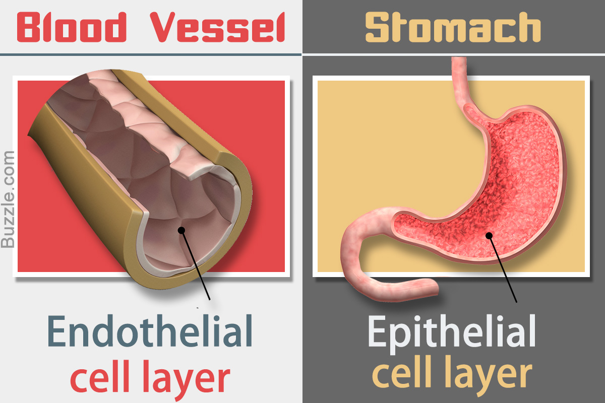
Endothelial cells line the interior of blood vessels, whereas the epithelial cells coat the inner surface of internal organs. The epithelial cells also cover the external surface of the body.
Did You Know?
Urinalysis showing a large number of epithelial cells in the urine may indicate a urinary tract infection.
Endothelial and epithelial cells that make up the tissues in humans and animals are of epithelial origin, but there is a major difference between the location, structure, and function of these cells. However, both the cells that form the tissue act like an interface between the underlying layer and the outside environment. Endothelial cells lie ‘inside’ the body as they are located in the blood vessels. On the other hand, epithelial cells are often described as lying ‘outside’ the body as they also coat the outer layer (epidermis) of the skin. The following BiologyWise article discusses the various aspects of these cells.
Endothelial Cells Vs. Epithelial Cells
Location
✦ Endothelial cells form the endothelium, a thin layer that coats the inner surface of blood vessels. Simply said, the cells always lie attached to the vessel wall. So, the inner wall of the entire circulatory system is covered with endothelial cells. These cells act as an interface between the circulating blood and the vessel wall. The endothelium is one-cell-layer thick and also lies attached to the interior of the heart chambers.
✦ Epithelial cells that form the epithelium not only cover the outside portion of the body, but also provide a coating to all the internal organs of the body. For instance, the outermost layer of the skin, the epidermis, is nothing but the epithelium. Thus, the superficial skin is lined by epithelial cells, thereby, forming a protective barrier against the external environment. The epithelial cells also line the inner surface of the liver, stomach, intestine, lungs, urethra, urinary bladder, and other organs of the body. In other words, epithelial cells provide a coating to the surface of the body as well as to its internal tissues.
Function
✦ The endothelial cells that lie attached to the vascular wall play a key role in controlling blood flow. The cells secrete nitric oxide (NO), which allows dilation of blood vessels to increase blood circulation. This also helps in controlling blood pressure. Endothelial cells also produce different kinds of proteins to keep blood clots at bay. However, it also releases clotting proteins to stop bleeding. Endothelial cells found in the glomeruli (capillaries located in the kidneys) also help in filtering blood.
✦ The epithelial cells, that form the skin, keep the underlying tissue safe from injury, bacterial invasion, exposure to hazardous chemicals, and excessive loss of moisture. The epithelial cells on the skin also release sweat whenever necessary, which helps in regulating body temperature. The epithelial cells that line the pancreas secrete enzymes to promote digestion. On the other hand, the epithelial cells that coat the small intestine help in absorbing nutrients from the ingested food. The epithelial cells that form the mucous membrane line the respiratory tract. The membrane secretes mucus, which traps inhaled bacteria and viruses and stops them from entering the lungs. Specialized epithelial cells located in the skin, nose, tongue, and eyes interact with the nerve endings, which help detect sensory stimuli. In summation, the epithelial cells are primarily involved in secretion, absorption, and protection.
Structure
✦ The endothelial cells that make up the endothelium are a single layer of cells. This single sheet of endothelium is so thin that molecules of water and oxygen can easily penetrate the layer and access the surrounding tissue. Also, the endothelial cells lack a tightly packed epithelial morphology. They are spaced by intercellular clefts that allow passage of fluids and diffusion of substances.
✦ The epithelial cells that form the epithelium provide multiple layers of protection from the outside environment. Epithelial cells form a closely packed structure similar to bricks stacked one above the other in a wall, with very little extracellular space between them.
Intermediate Filaments
✦ Certain proteins, referred to as intermediate filaments, assist in providing support and defining the shape of cells. In short, they provide structure to cells. In case of endothelial cells, vimentin filaments provide structure and shape to the cells.
✦ Keratin filaments play a crucial role in determining the epithelial cell structure.
Surface Layer
✦ Another major difference lies in their surfaces. The endothelial cells that make up the endothelium has a non-thrombogenic smooth and soft surface, meaning blood does not clot during normal circulation.
✦ The epithelium is composed of different types of epithelial cells, so the surface layer is seen having irregular papillary projections.
Disclaimer: The information provided in this article is solely for educating the reader. It is not intended to be a substitute for the advice of a medical expert.

