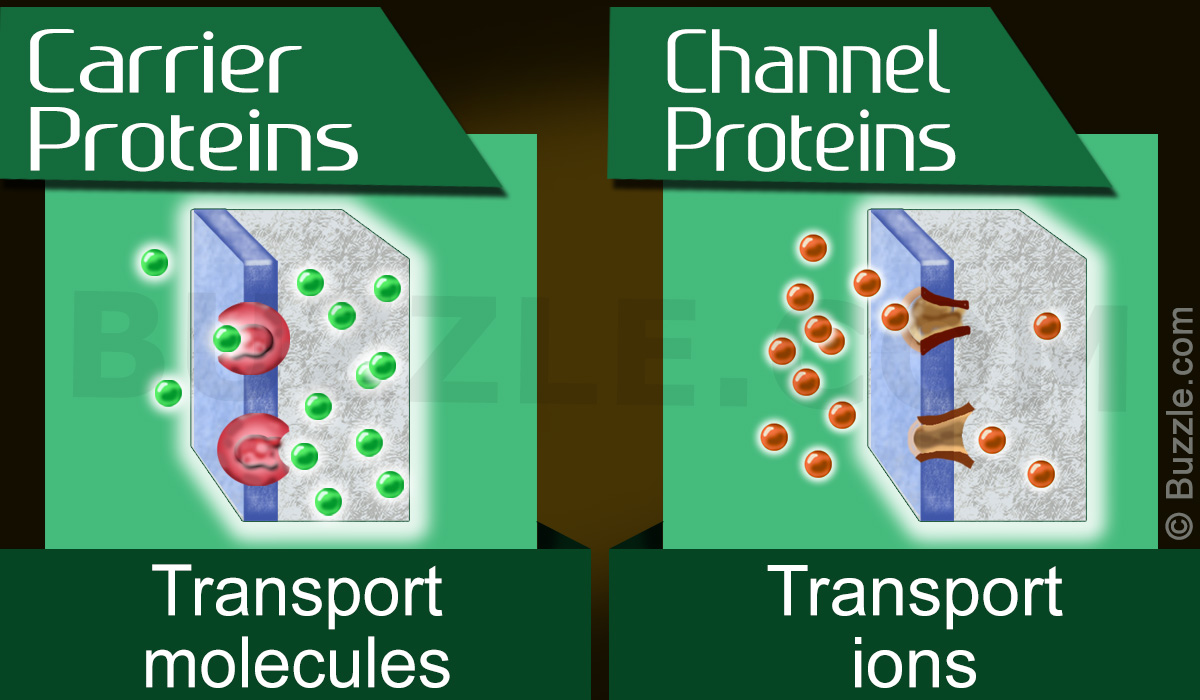
The proteins that facilitate the movement of molecules across a biological membrane are transport proteins. Carrier proteins and channel proteins are two types of membrane proteins. Here, we do an analysis of carrier proteins vs. channel proteins for a better understanding of the same.
Did You Know?
The Na+/K+ pump was discovered by Jens Christian Skou in the nerve cell of crabs in the year 1957 and was awarded the Noble Prize in Chemistry in 1997.
Biological membranes contain an impermeable lipid bilayer with selectively permeable proteins embedded in it. The transport of ions and small molecules across a biological membrane is called membrane transport. These biological membranes regulate the traffic of molecules that may enter or leave the cell. Transport of certain small, uncharged molecules like O2, CO2, and urea across the cell membrane may take place though slowly, but readily on its own.
Proteins that function in the transport of solutes across biological membranes are called membrane transport proteins. They hasten the rate at which molecules are transported across the biological membrane. These are membrane integral proteins (span across the membrane) and are highly specific in nature (one type of protein interacts with only one type of solute molecules).
Carrier Proteins
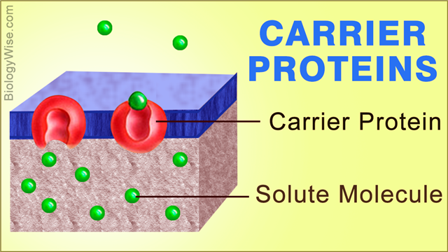
The function of carrier proteins is to transfer a large number of both polar and non-polar molecules across the semipermeable biological membrane. Carrier proteins exist in two conformations: (i) conformation A – the binding site is empty and; (ii) conformation B – the binding site is occupied by the solute. They possess a specific binding site to which the solute molecules bind. This binding of the solute molecules causes the protein to change its conformation to B, such that the binding site of the protein is now exposed on the other side of the membrane. The solute is then released from the carrier protein, and it returns to its original conformation A.
Channel Proteins
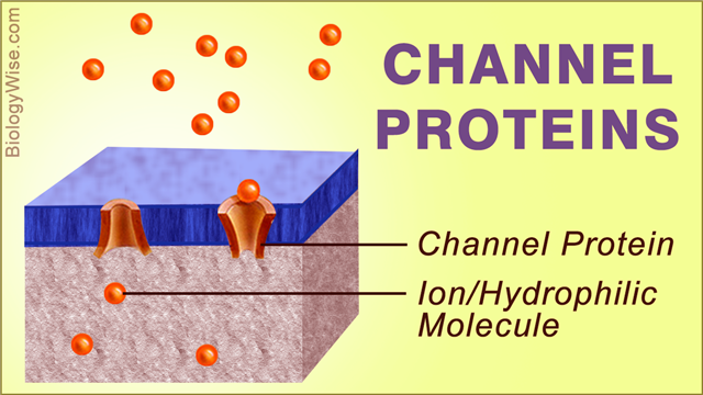
The function of channel proteins is to transfer water molecules and small polar molecules across the semipermeable biological membrane. They form a water-filled passage made of hydrophilic proteins that help in the transfer of ions and small polar solutes across the biological membrane. They are basically a pore whose walls are made of proteins.
Similarities Between Carrier and Channel Proteins
- Both proteins hasten the rate of transfer of molecules across the biological membranes.
- They span across the biological membrane.
- They are highly specific for the molecules they transfer.
- In case of both, the rate of reaction increases with an increase in ion concentration, till it reaches saturation after which, the rate remains steady even with an increase in the concentration of the solute.
Differences Between Carrier and Channel Proteins
Mechanism
▶ Carrier proteins transfer solutes across the biological membrane by binding to the solute and alternate between two conformations. Their mechanism is similar to enzyme-substrate reactions following Michaelis-Menten equation (however, they do not change the substrate, i.e., the solute).
▶ Channel proteins interact the least with the solute they transfer.
Nature of Solute
▶ Carrier proteins transfer both polar and nonpolar solutes across the biological membrane.
▶ Channel proteins transfer only small and polar solutes across the biological membrane.
Specificity
▶ Specificity of carrier proteins is due to the specific binding sites to which the solute molecules bind.
▶ Specificity of channel proteins is due to an ion selectivity filter.
Ion selectivity filter: In simplest words, it can be defined as the narrowest part of the pore which will only allow the passage of specific molecules with a particular size and charge to pass through.
Rate of Transfer of Solute
▶ The rate of transfer of solute by carrier proteins is about 104 ions per second.
▶ As the proteins do not flip from one conformation to the other, the rate of transfer of solute by channel proteins is much higher, i.e., 108 ions per second.
Nature of Transport
▶ Carrier proteins usually transport molecules against the concentration gradient; to do so, they require energy. This energy can be supplied to it either by hydrolysis of ATP (known as active transport) or can be coupled with the transfer of another solute molecule (known as facilitated diffusion).
Examples of Carrier Protein-mediated Active Transport
1. Na+/K+ ATPase: It plays an important role in the uptake of glucose by the cell. Three Na+ ions are pumped out of the cell, and two K+ ions are pumped inside the cell. This takes place against the concentration gradient, and 1 ATP is consumed for this process. These Na+ ions help to bring glucose inside the cell (discussed below).
2. SR Ca2+ ATPase: It is present in the Sarcoplasmic Reticulum (abbreviated as SR; specialized Endoplasmic Reticulum present in muscle cells). Two Ca2+ ions are transported from the cytosol into the SR. This step consumes 1 ATP and is required for the contraction of muscles.
Examples of Carrier Protein-mediated Facilitated Diffusion
1. Symport: Co-transport of molecules or ions in the same direction of the biological membrane.
E.g. Na+-driven glucose pump: It is present in the intestinal epithelial cells. Glucose is taken up by the intestinal cells with this pump. Here, the glucose moves against its concentration gradient. The energy to perform this is supplied by the movement of Na+ ions into the cells; this movement is down its concentration gradient and is favored.
2. Antiport: Co-transport of molecules or ions in the opposite direction of the biological membrane.
E.g. Na+/Ca2+ exchanger: Here, Na+ ions move down their concentration gradients, and this provides energy for the movement of Ca2+ against its concentration gradient. Three Na+ ions are transported inside the cell, and one Ca2+ ion is pumped out of the cell.
Channel proteins always transport molecules down the concentration gradient by a process of diffusion and, hence, mediate passive transport.
Examples of Channel Protein-mediated Passive Transport
Ion channels are not continuously open and are said to be gated, which open only in response to specific stimulus.
1. Voltage-gated Channels: These ion channels are activated when there is a potential difference generated across the biological membrane. An example is voltage-dependent calcium channels, which are found on the cell membrane of neurons, glial cells, and muscle cells. These channels are activated when there is a potential difference across the membrane and cause an influx of Ca2+ ions inside the cells. They may play a role in neurotransmission, muscle relaxation, gene expression, etc., depending on the type of cell on which they are present.
2. Ligand-gated Channels: Nicotinic Acetylcholine Receptor (nAchR) channel is usually found in neuromuscular junctions. When the nAchR channel binds to the neurotransmitter, acetylcholine (ligand), the closed channels open and allow the influx of Na+ ions, thus helping in the contraction of muscles.

